Innovative kinome activity technology


Our multiplex kinase activity profiling technology reveals the bigger picture of cellular signaling and provides mechanistic insights needed to fully understand diseases and cell alterations. It opens up opportunities for a wide range of rapid and non-invasive applications and is applied in the areas of fundamental science, translational research and diagnostics.

Innovative kinome activity technology


Our multiplex kinase activity profiling technology reveals the bigger picture of cellular signaling and provides mechanistic insights needed to fully understand diseases and cell alterations. It opens up opportunities for a wide range of rapid and non-invasive applications and is applied in the areas of fundamental science, translational research and diagnostics

Kinases are a protein family playing a critical role in signal transduction which underlie many cellular processes. PamGene’s functional kinase assay is activity-based and detects protein kinase activity directly in cellular and tissue lysates, through measuring peptide phosphorylation by protein kinases.

Our technology opens up opportunities for a wide range of rapid and non-invasive applications. It is currently applied in the areas of fundamental science, translational research and diagnostics. PamGene’s technology is used to gain insights into discriminative drug responses, analyzing and optimizing kinase inhibitors and discovery of novel biomarkers, targets and pathways.

The PamChip®


The PamChip is available with either phosphotyrosine kinase (PTK) or serine-threonine kinase (STK) arrays and reagents. The PamChip® contains 4 identical peptide arrays, each array containing 144 (STK) or 196 (PTK) peptide sequences 13 amino acids long. The peptides harbor phosphorylation sites derived from literature or computational predictions and are correlated with one or multiple upstream kinases (Protein tyrosine kinases for the PTK PamChip® and Serine-threonine kinases for the STK PamChip®).

The surface of the PamChip® microarrays is three dimensional (3D), composed of aluminum oxide, which has a porous structure with long branched interconnected capillaries having a diameter of about 200 nm. The reduced distance of the target and probe molecules allows for faster, diffusion-independent protein binding and enzymatic reactions. The 3D surface of the PamChip is designed to allow peptide substrates to be deposited at higher concentrations than conventional arrays, increasing the sensitivity.


The PamChip


The PamChip is available with either phosphotyrosine kinase (PTK) or serine-threonine kinase (STK) arrays and reagents. The PamChip® contains 4 identical peptide arrays, each array containing 144 (STK) or 196 (PTK) peptide sequences 13 amino acids long. The peptides harbor phosphorylation sites derived from literature or computational predictions and are correlated with one or multiple upstream kinases (Protein tyrosine kinases for the PTK PamChip® and Serine-threonine kinases for the STK PamChip®).

The surface of the PamChip® microarrays is three dimensional (3D), composed of aluminum oxide, which has a porous structure with long branched interconnected capillaries having a diameter of about 200 nm. The reduced distance of the target and probe molecules allows for faster, diffusion-independent protein binding and enzymatic reactions. The 3D surface of the PamChip is designed to allow peptide substrates to be deposited at higher concentrations than conventional arrays, increasing the sensitivity.


PamChip Assay Principle

 
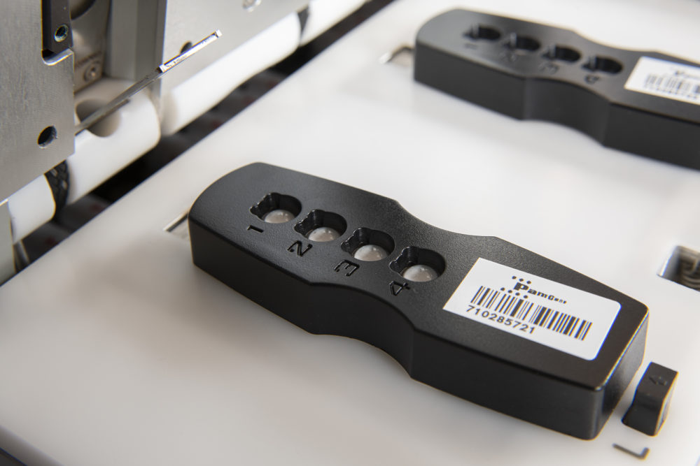
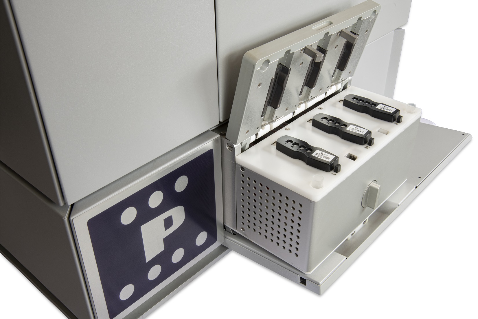
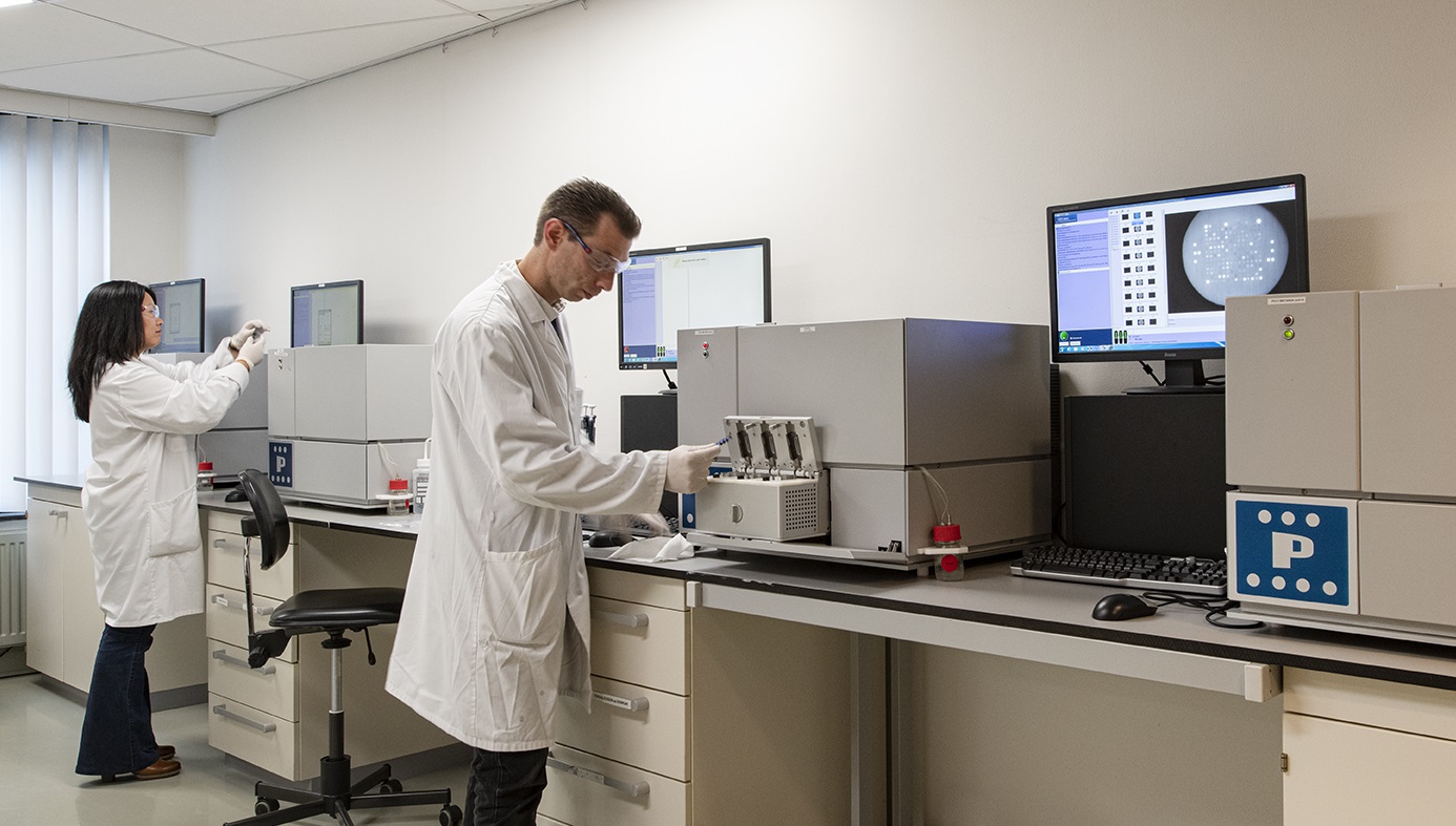
Fluorescently labelled anti-phospho antibodies are used to detect phosphorylation activity of kinases present in the sample. The sample consisting of protein and labelled antibodies are dispensed onto the PamChip, which is placed in the PamStation. Once inside the instrument the sample is incubated at a predetermined temperature. During incubation the sample is pumped back and forth through the porous material to maximize binding kinetics and minimize assay time. Reaction times are typically minutes to an hour. Upon wetting, the surface material of the PamChip microarrays becomes translucent, facilitating imaging using a LED imaging system.

PTK Assay

Kinase(s) in the sample (recombinant, cell or tissue lysate) actively phosphorylate substrates on the PamChip®, in the presence of ATP. A FITC-conjugated PY20 antibody, present in the reaction mix is used to detect the phosphorylated Tyr to quantify the phosphorylation signal. Images are taken every 5 minutes to generate real time kinetics data.

STK Assay

Kinase(s) in the sample (recombinant, cell or tissue lysate) actively phosphorylate substrates on the PamChip®, in the presence of ATP. An antibody mix is used to detect the phosphorylated Ser/Thr, and a 2nd FITC-conjugated antibody is used in a detection mix to quantify the phosphorylation signal.

The PamStation


The PamStation is a fully automated instrument designed for processing PamChip microarrays. 3 PamChips can be processed simultaneously on the PamStation. It is controlled by a dedicated computer with access to advanced software for data analysis, called the BioNavigator. The instrument is CE marked.

Key attributes:

  • Fully integrated incubation, washing and image acquisition.
  • Dimensions are 49cm (height) x 66cm (width) x 58cm (depth), weight is 82 kgs.
  • Instrument is controlled by a dedicated computer and software (Evolve; included).
  • Real-time detection offers unique capability of generating kinetic data on enzyme/substrate interactions.
  • Easy to operate, very limited maintenance required.

BioNavigator


BioNavigator is PamGene’s powerful software tool for interpretation of PamChip measurements with its capabilities including: Visualization, Computation, Storage and Sharing.

BioNavigator is the unique tool for performing data analysis for many types of PamChip experiments. It treats data analysis as a connection of modular series of data transformation steps, called a data analysis protocol. BioNavigator creates and executes data analysis protocols. There is the choice of either using pre-existent standard data analysis protocols or creating customized versions.

Products and services


We offer Contract Research Services tailored to your wishes, needs and research questions. It is also possible to obtain your own PamStation. The PamGene team will support you in making the PamStation an integral part of your research facility’s departments by providing in-house training and support.

Scientific background

The active kinome: The modern view of how active protein kinase networks fit in biological research

Khaled Alganem1, Robert McCullumsmith et al.

www.sciencedirect.com Current Opinion in Pharmacology (2022)

Publication

Subcellular partitioning of protein kinase activity revealed by functional kinome profling

LaurenWegman‑Points1, Li‑LianYuan

Scientific Reports | (2022) 

Publications

Oxidative stress-induced FAK activation contributes to uterine serous carcinoma aggressiveness

Isabel C. Lopez-Mejia, Andree Yeramian et al,

Molecular Oncology 17 (2023)

Publication